|
Regarding the concept that a medieval artist might have produced such an image, Schumacher said: "How and why would an artist embed three dimensional information in the gray shading of an image when no means of viewing this property would be available for at least 650 years after it was done...and how could the artist control the quality of the work when the gray scale could not be seen as elevation?"
To date the only technique that has come anywhere near giving the shroud's 3-D effect with a VP-8 analyzer comes from an experiment carried out by Dr August Accetta, a medical practitioner with much experience in the use of autoradiography in diagnostic medicine. For this he used himself as guinea pig, injecting himself with a suitable dose of radioactive methylene diphosphate. With the aid of his wife, he then used a gamma camera to capture the photons derived from gamma radiation coming from his body. The image so obtained was then scanned with the VP-8 Analyzer. The result showed a 3-D effect similar in major details to what was obtained from the Turin shroud but sadly lacking in the fine detail.
This was Dr Accetta's first trial and no doubt could be improved. It definitely establishes that exposure to radiation can somehow be linked to this 3-D image effect.
Further evidence that is contrary to the Turin shroud being of medieval and European origin is present on samples taken with small strips of sticky tape pressed against the cloth's surface. This yields copious amounts of plant debris thought to have come from people having placed flowers upon the shroud. Among the debris, professor, Avinoam Danim from the Hebrew University in Jerusalem, an acknowledged expert in the flora of Israel, has identified pollen from three species that grow together only in a twenty mile region between Jerusalem and Hebron.
Of these three, one is Cistus creticus, another Zygophylum dumosum which grows only in Israel, Jordan, and the Sinai, and a third, Gundelia tourneforte, that is distinctively Middle East and absent from Europe and flowers only between March and May, the period in which Jesus was crucified.
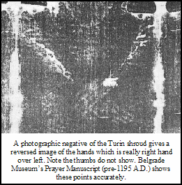
Assertions for the shroud being a fake have claimed it is either a painting or else that some unknown person in medieval times invented a pinhole camera plus a method for developing the resulting image on a very large sheet. There is a wealth of evidence against either hypothesis, but little need to repeat it in the face of the 3-D effects obtained with the shroud and NASA's VP-8 image analyzer. The facts are that nobody has been able to simulate the image on the shroud and retain all its characteristics, not even with the most modern technology.
The bloodstains too, are a thorn in the sides of those who cry fake. Recent work has shown the blood and serum goes right through the cloth of the shroud to the other side, something that does not happen with the medieval technique of brushing on iron oxide powder in a gelatin protein base and enhancing the blood color with cinnabar. The shroud's blood has been analyzed with modern technology and shown to be type A-B. Also demonstrated are DNA sequences that specify genes for both the X and the Y chromosomes, thereby identifying the blood as originating from a male person.
A key question remaining is the method by which the image was transferred to the cloth. The Urantia Book informs us that dissolution of Jesus' body took place by the natural mode except it was greatly accelerated. The natural mode is microbial-induced decomposition, the final product being carbon dioxide and water--but let's not forget the bones. Bones do not oxidize in this way. Normally bone decomposition is exceedingly slow dissolution through attack by soil acids. So how was the acceleration of bone and flesh decomposition achieved?
People interested in how an image could be transferred to the linen of the Turin shroud have suggested radiation-induced dematerialization of the body of Jesus may have been the cause, one suggesting neutron bombardment2, while another suggests "weak dematerialization" associated with spontaneous pion (pi-meson) decay3.
Perhaps the reality was that some form of radiation was utilized to accelerate the normal decay process? If so and if radiation was required to give the 3-D information that is contained in the shroud image, then perhaps it started soon after Jesus' body became lifeless, then continued at a slow pace that did not result in excessive heat generation over about 36 hours of entombment prior to the women arriving at the tomb early on Sunday morning.
|
|


